Eyelid anatomy, illustrated
The layers of the upper and lower eyelid, the fat pads behind them and how the main operations reach them. Each illustration is drawn from public anatomy plates and checked against the anatomy literature; the numbers match the key beside it.
The eyelid, layer by layer
Medical illustrations, labelled. The numbers match the key.
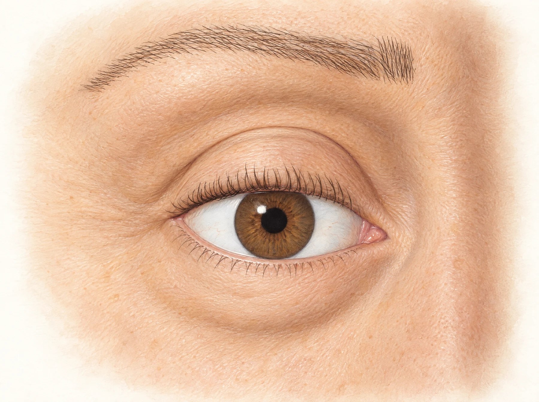
Eyelid surface landmarks
The right eye of a woman in her mid-fifties, showing the brow, the upper lid crease and fold, the corners of the eye, the tear trough and the lid-cheek junction.
- Eyebrow
- Upper eyelid crease
- Upper eyelid skin fold
- Upper eyelid margin and lashes
- Lateral canthus
- Medial canthus
- Caruncle
- Lower eyelid margin
- Lower eyelid
- Tear trough
- Lid-cheek junction
- Cheek
- Iris
- Pupil
- Sclera
Anatomy checked against StatPearls. Anatomy, Head and Neck: Eyelid, StatPearls. Blepharoplasty, StatPearls. Blepharoplasty, Lower Lid, Canthal Support, Wong CH et al. The tear trough ligament: anatomical basis for the tear trough deformity. Plast Reconstr Surg 2012, Muzaffar AR et al. Surgical anatomy of the ligamentous attachments of the lower lid and lateral canthus. Plast Reconstr Surg 2002.
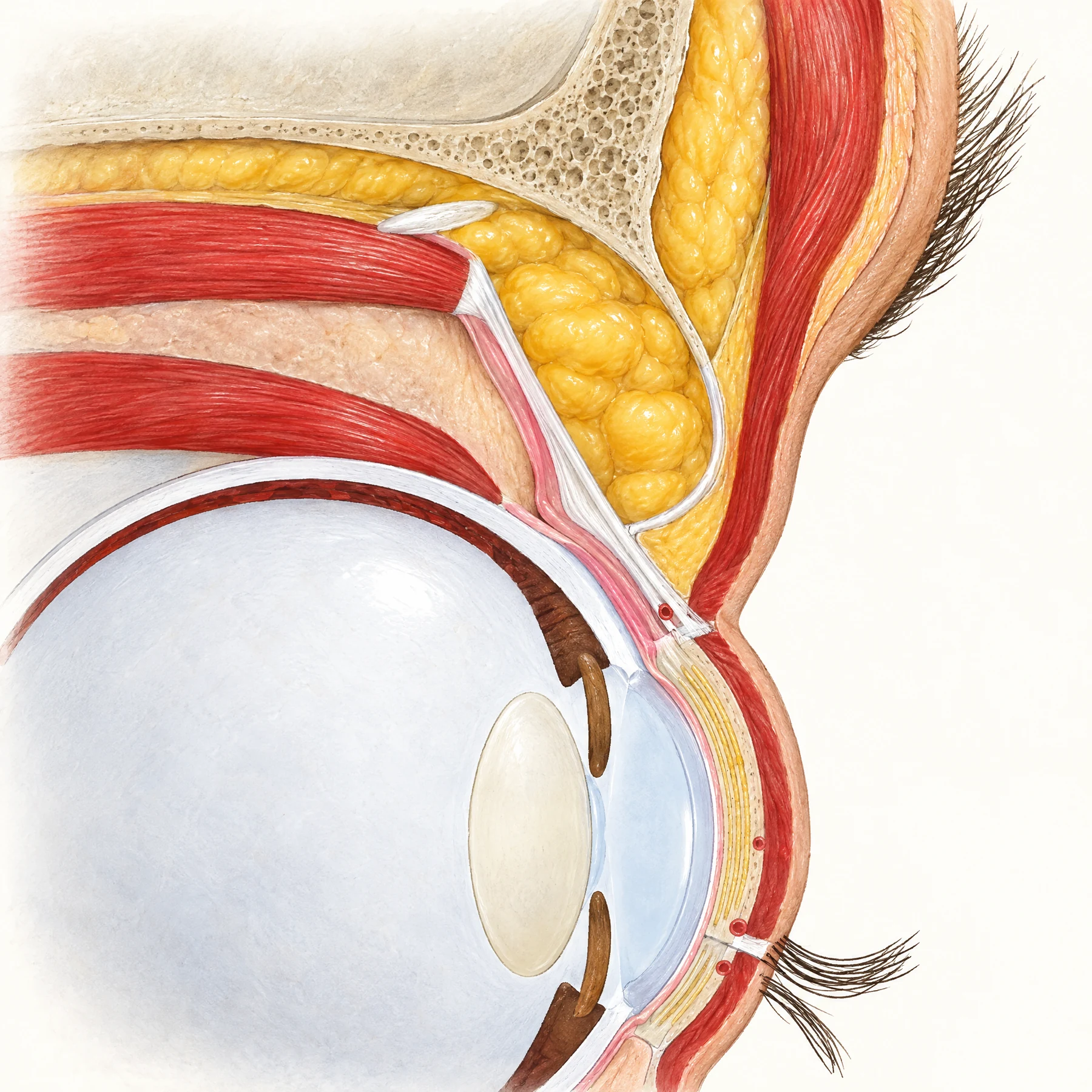
Upper eyelid cross-section
Cross-section of the closed upper eyelid through the middle of the pupil, from the brow down to the lashes.
- Eyebrow
- Orbicularis oculi, orbital part
- Brow fat (ROOF)
- Frontal bone, superior orbital rim
- Orbital septum
- Preaponeurotic fat pad
- Whitnall ligament
- Levator palpebrae superioris muscle
- Levator aponeurosis
- Müller muscle
- Superior rectus muscle
- Superior conjunctival fornix
- Orbicularis oculi, preseptal part
- Peripheral arterial arcade
- Eyelid crease
- Tarsus with meibomian glands
- Orbicularis oculi, pretarsal part
- Skin
- Eyelashes
- Cornea
- Iris
- Lens
Anatomy checked against StatPearls. Anatomy, Head and Neck: Eyelid, StatPearls. Anatomy, Head and Neck: Eye, Superior Tarsal Muscle (Müller Muscle), Hwang K. Surgical anatomy of the upper eyelid relating to upper blepharoplasty or blepharoptosis surgery. Anat Cell Biol 2013, Meyer DR et al. Anatomy of the orbital septum and associated eyelid connective tissues. Ophthalmic Plast Reconstr Surg 1991, Anderson RL, Beard C. The levator aponeurosis. Attachments and their clinical significance. Arch Ophthalmol 1977, Kakizaki H et al. Whitnall ligament anatomy revisited. Clin Exp Ophthalmol 2011, May JW Jr et al. Retro-orbicularis oculus fat (ROOF) resection in aesthetic blepharoplasty. Plast Reconstr Surg 1990, EyeWiki. Aponeurotic Blepharoptosis, Gray's Anatomy (1918), fig. 893: sagittal section of the upper eyelid.
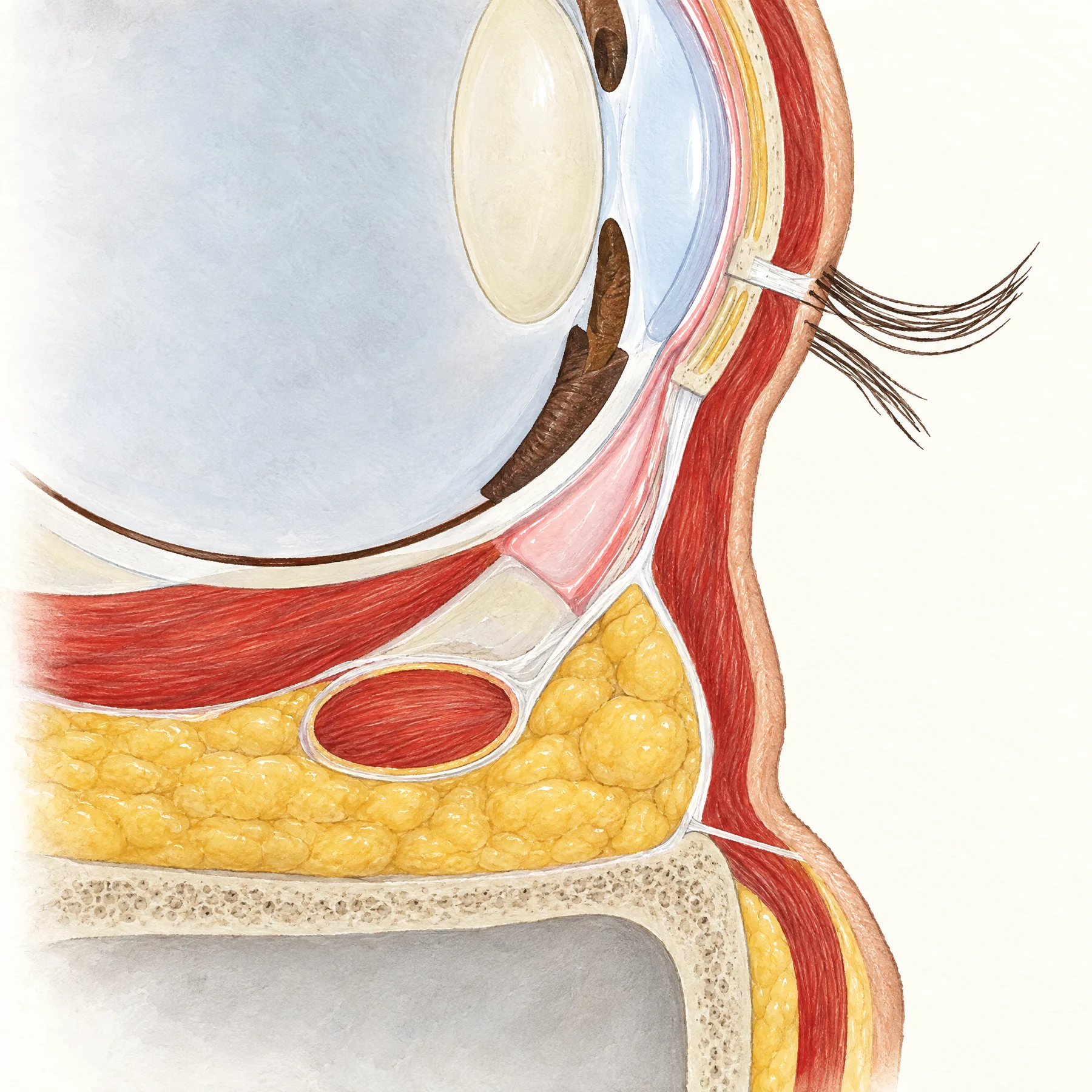
Lower eyelid cross-section
Cross-section of the closed lower eyelid through the middle of the pupil, from the lashes down to the cheek.
- Skin
- Orbicularis oculi, pretarsal part
- Tarsus with meibomian glands
- Eyelashes
- Orbicularis oculi, preseptal part
- Capsulopalpebral fascia (lower lid retractor)
- Orbital septum
- Central fat pad
- Conjunctiva
- Inferior conjunctival fornix
- Inferior oblique muscle
- Inferior rectus muscle
- Orbital fat
- Orbital floor (maxilla)
- Maxillary sinus
- Inferior orbital rim
- Orbicularis retaining ligament
- Lid-cheek junction
- Suborbicularis oculi fat (SOOF)
- Orbicularis oculi, orbital part
- Subcutaneous fat
- Cornea
- Iris
- Lens
- Upper eyelid
Anatomy checked against StatPearls. Anatomy, Head and Neck: Eyelid, Hwang K. Surgical anatomy of the lower eyelid relating to lower blepharoplasty. Anat Cell Biol 2010, Hawes MJ, Dortzbach RK. The microscopic anatomy of the lower eyelid retractors. Arch Ophthalmol 1982, Kakizaki H et al. Lower eyelid anatomy: an update. Ann Plast Surg 2009, Kawakita T et al. Measurement of fornix depth and area. Eye 2009, Muzaffar AR et al. Surgical anatomy of the ligamentous attachments of the lower lid and lateral canthus. Plast Reconstr Surg 2002, Aiache AE, Ramirez OH. The suborbicularis oculi fat pads: an anatomic and clinical study. Plast Reconstr Surg 1995, Gray's Anatomy (1918), fig. 891: sagittal section of the orbit.
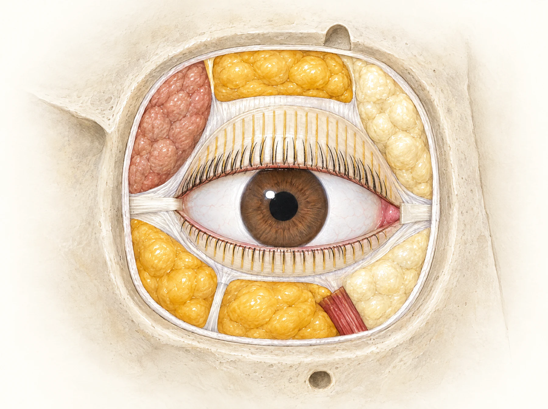
Orbital fat pads, front view
The right eye from the front with the skin and orbicularis removed and the orbital septum opened, showing the fat pads of the upper and lower eyelids.
- Lacrimal gland
- Central fat pad, upper eyelid
- Nasal fat pad, upper eyelid
- Levator aponeurosis
- Upper tarsus with meibomian glands
- Lateral canthal tendon
- Medial canthal tendon
- Caruncle
- Lower tarsus
- Lateral fat pad, lower eyelid
- Central fat pad, lower eyelid
- Medial fat pad, lower eyelid
- Inferior oblique muscle
- Arcuate expansion
- Cut edge of the orbital septum
- Supraorbital notch
- Infraorbital foramen
- Lateral orbital rim
- Iris
- Sclera
Anatomy checked against StatPearls. Anatomy, Head and Neck: Eyelid, StatPearls. Blepharoplasty, StatPearls. Transconjunctival Blepharoplasty, Hwang K. Surgical anatomy of the lower eyelid relating to lower blepharoplasty. Anat Cell Biol 2010, Massry GG. Prevalence of lacrimal gland prolapse in the functional blepharoplasty population. Ophthalmic Plast Reconstr Surg 2011, Gray's Anatomy (1918), fig. 894: tarsi and their ligaments.
How the operations are done
Medical illustrations, labelled. The numbers match the key.
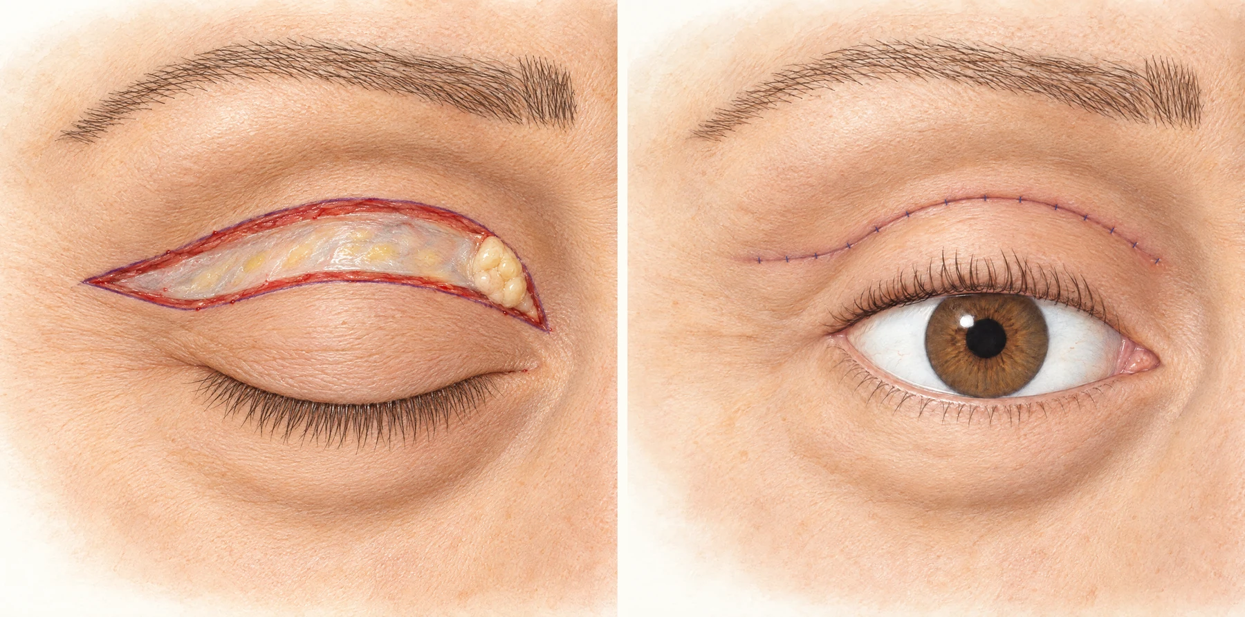
Upper blepharoplasty
Upper eyelid skin is removed through an incision in the natural crease (left), and the wound is closed so the scar lies in the crease (right).
- Lower incision line in the eyelid crease
- Upper incision line
- Orbital septum
- Central fat pad behind the septum
- Nasal fat pad
- Orbicularis oculi, cut edge
- Lateral end of the incision
- Closed incision in the eyelid crease
- Lateral end of the closed incision
Anatomy checked against StatPearls. Blepharoplasty, StatPearls. Anatomy, Head and Neck: Eyelid, Hwang K. Surgical anatomy of the upper eyelid relating to upper blepharoplasty or blepharoptosis surgery. Anat Cell Biol 2013.
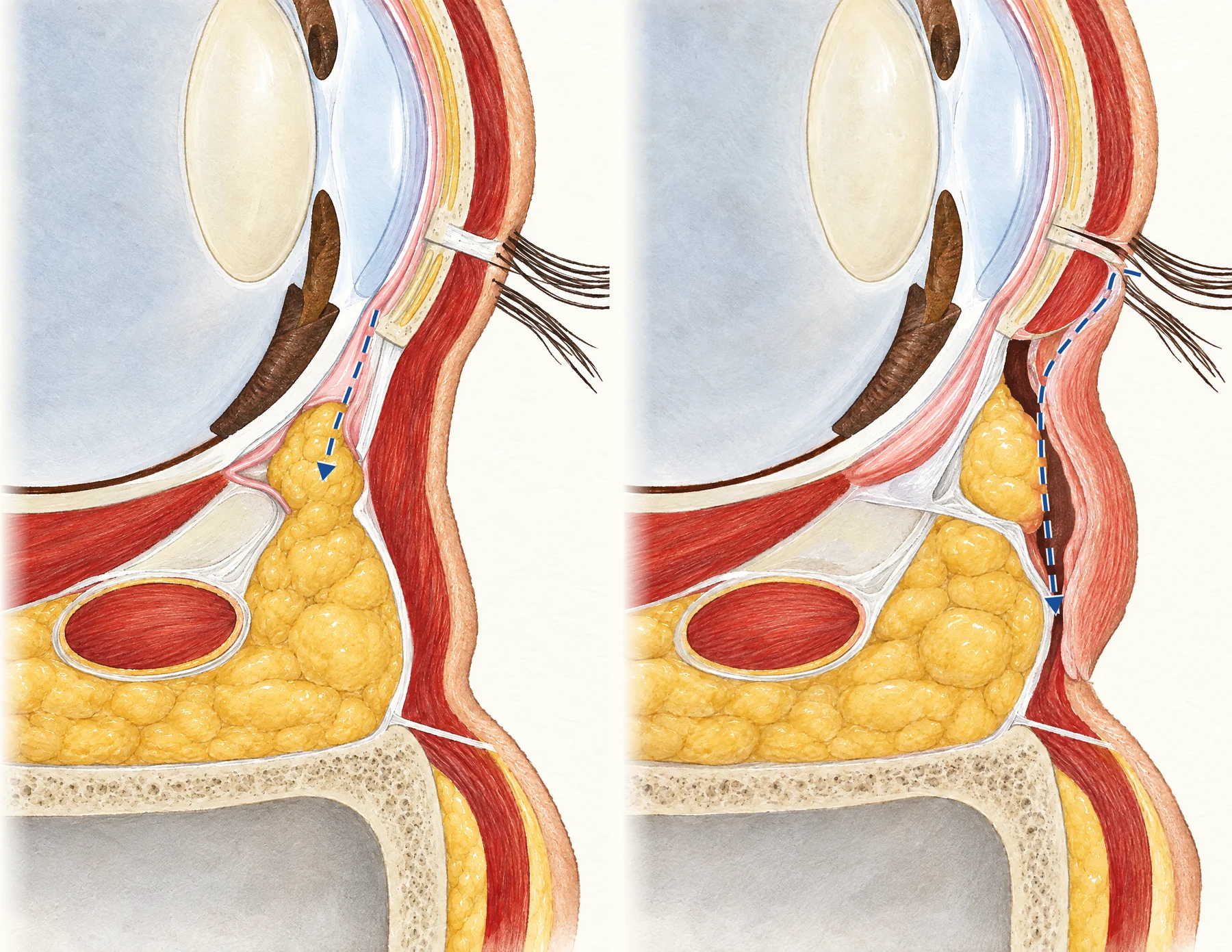
Lower blepharoplasty approaches
The lower eyelid fat is reached either through the inside of the lid (left, transconjunctival) or through a skin incision just below the lashes (right, subciliary).
- Transconjunctival incision
- Central fat pad
- Orbital septum, not opened
- Skin and orbicularis, not cut
- Lower tarsus
- Inferior oblique muscle
- Subciliary skin incision
- Pretarsal orbicularis, preserved
- Skin-muscle flap
- Plane in front of the septum
- Orbital septum, opened
- Central fat pad
Anatomy checked against StatPearls. Transconjunctival Blepharoplasty, StatPearls. Blepharoplasty Subciliary Approach, StatPearls. Anatomy, Head and Neck: Eyelid, Hwang K. Surgical anatomy of the lower eyelid relating to lower blepharoplasty. Anat Cell Biol 2010.
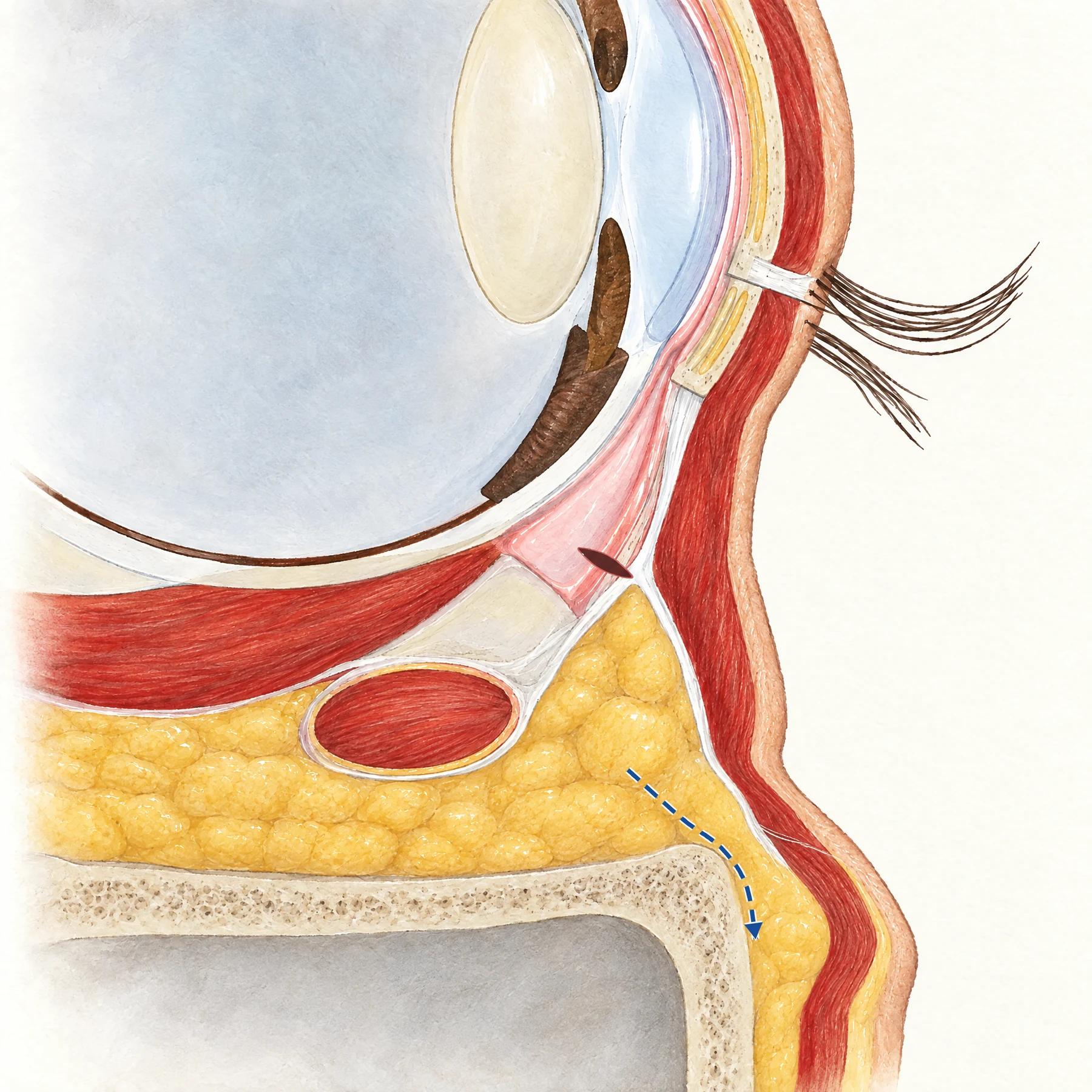
Fat repositioning
Cross-section of the lower eyelid after fat repositioning: through an incision inside the lid, the orbital septum is released from the bony rim and a tongue of eyelid fat, still attached at its base, is moved over the rim onto the cheekbone.
- Transconjunctival incision
- Central fat pad
- Fat moved over the orbital rim
- Orbital septum, released from the rim
- Inferior orbital rim
- Maxilla
- Orbicularis oculi muscle
- Orbicularis retaining ligament
- Skin
- Lower tarsus
- Inferior oblique muscle
- Conjunctiva
- Maxillary sinus
Anatomy checked against Hamra ST. Arcus marginalis release and orbital fat preservation in midface rejuvenation. Plast Reconstr Surg 1995, Goldberg RA. Transconjunctival orbital fat repositioning: transposition of orbital fat pedicles into a subperiosteal pocket. Plast Reconstr Surg 2000, StatPearls. Transconjunctival Blepharoplasty, Hwang K. Surgical anatomy of the lower eyelid relating to lower blepharoplasty. Anat Cell Biol 2010, Muzaffar AR et al. Surgical anatomy of the ligamentous attachments of the lower lid and lateral canthus. Plast Reconstr Surg 2002.
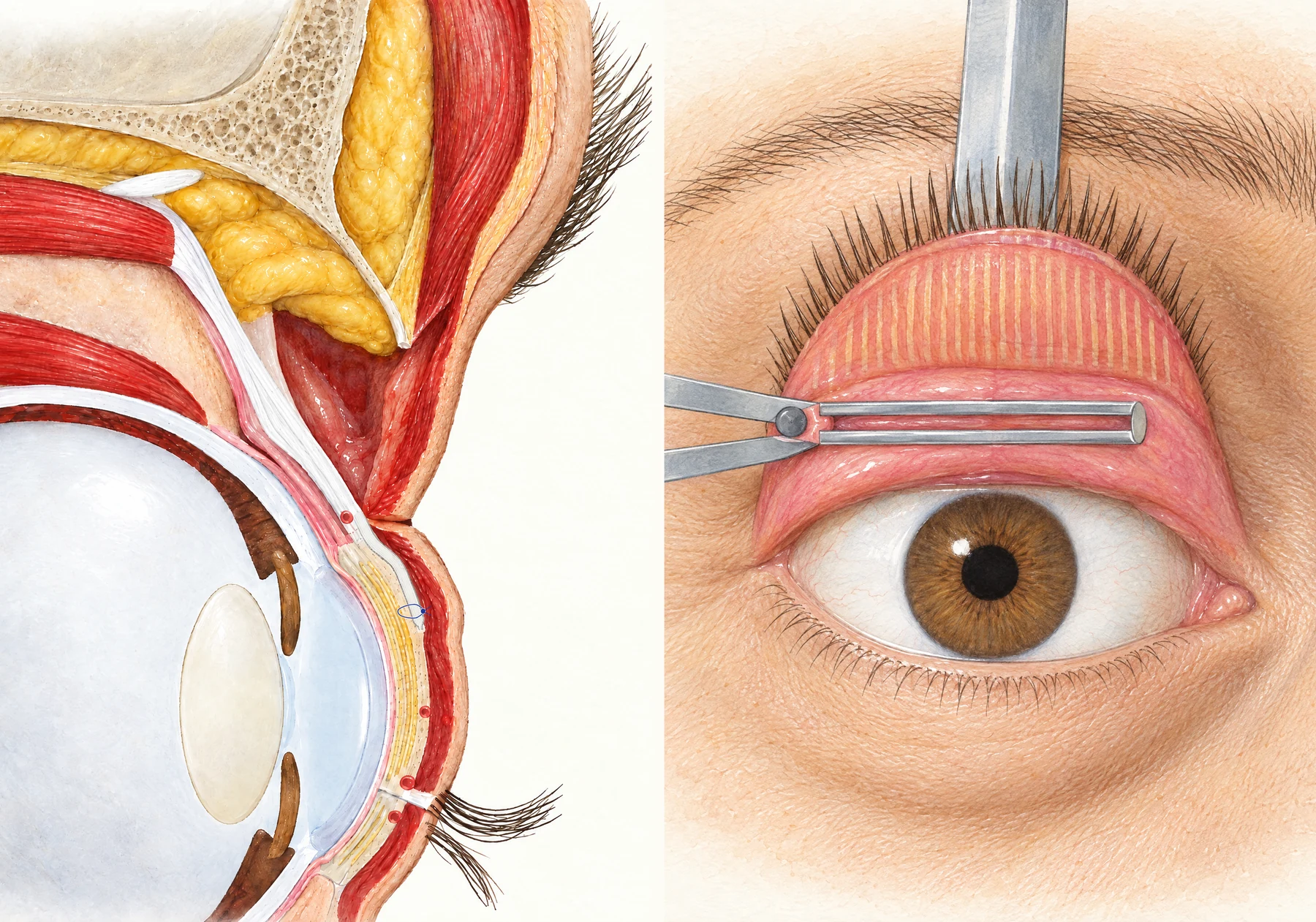
Ptosis repair
Two ways to raise a drooping upper eyelid: through a crease incision the levator aponeurosis is stitched to the front of the tarsus (left), or from inside the turned-out eyelid a strip of Müller muscle and conjunctiva is clamped for removal (right).
- Levator palpebrae superioris muscle
- Levator aponeurosis
- Müller muscle
- Preaponeurotic fat, lifted
- Orbital septum, opened
- Crease incision
- Orbicularis oculi muscle
- Tarsus
- Suture through the aponeurosis and the front of the tarsus
- Desmarres retractor
- Eyelashes of the everted eyelid
- Tarsus, inner surface
- Upper border of the tarsus
- Putterman clamp
- Müller muscle and conjunctiva in the clamp
Anatomy checked against EyeWiki. Aponeurotic Blepharoptosis, Anderson RL, Beard C. The levator aponeurosis. Attachments and their clinical significance. Arch Ophthalmol 1977, EyeWiki. Müller's Muscle-Conjunctival Resection (MMCR) Blepharoptosis Repair, Putterman AM, Urist MJ. Müller muscle-conjunctiva resection. Arch Ophthalmol 1975, StatPearls. Anatomy, Head and Neck: Eye, Superior Tarsal Muscle (Müller Muscle), StatPearls. Anatomy, Head and Neck: Eyelid, Hwang K. Surgical anatomy of the upper eyelid relating to upper blepharoplasty or blepharoptosis surgery. Anat Cell Biol 2013.
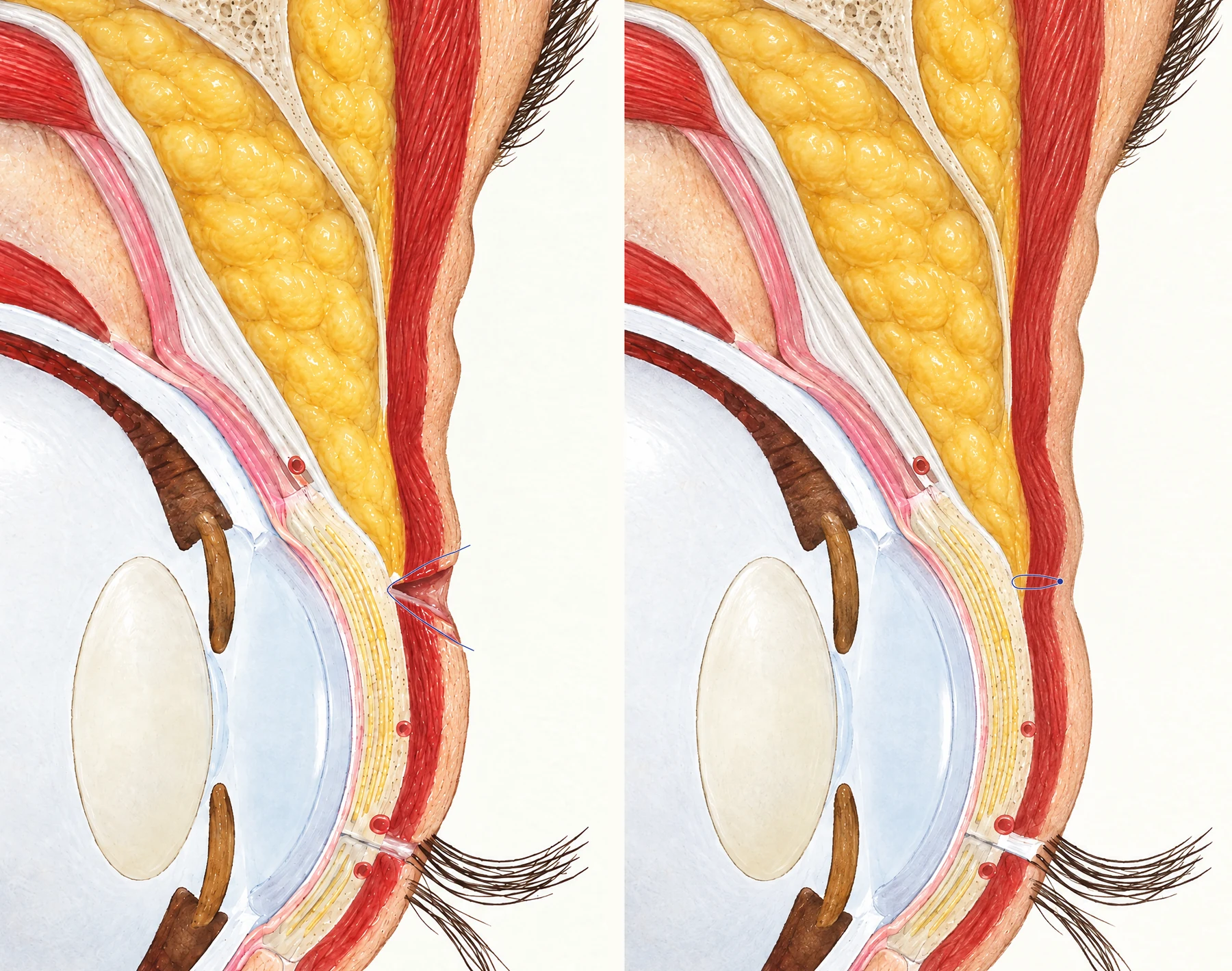
Double eyelid surgery
Cross-sections of an East Asian upper eyelid, where the septum and fat reach down in front of the tarsus. Left: incisional method, with a strip of tissue removed at the planned crease and a stitch joining the skin edges to the levator aponeurosis. Right: buried-suture method, with a stitch from under the skin to the tarsus and no incision.
- Levator aponeurosis
- Müller muscle
- Preaponeurotic fat
- Orbital septum
- Orbicularis oculi muscle
- Tarsus
- Incision at the planned crease
- Suture from the skin edges to the levator aponeurosis
- Buried suture, knot under the skin
- Fat in front of the upper tarsus
- Septum joining the aponeurosis on the tarsus
Anatomy checked against EyeWiki. Asian Blepharoplasty (Double Eyelid Procedure), Jeong S et al. The Asian upper eyelid: an anatomical study with comparison to the Caucasian eyelid. Arch Ophthalmol 1999, StatPearls. Anatomy, Head and Neck: Eyelid, Hwang K. Surgical anatomy of the upper eyelid relating to upper blepharoplasty or blepharoptosis surgery. Anat Cell Biol 2013.
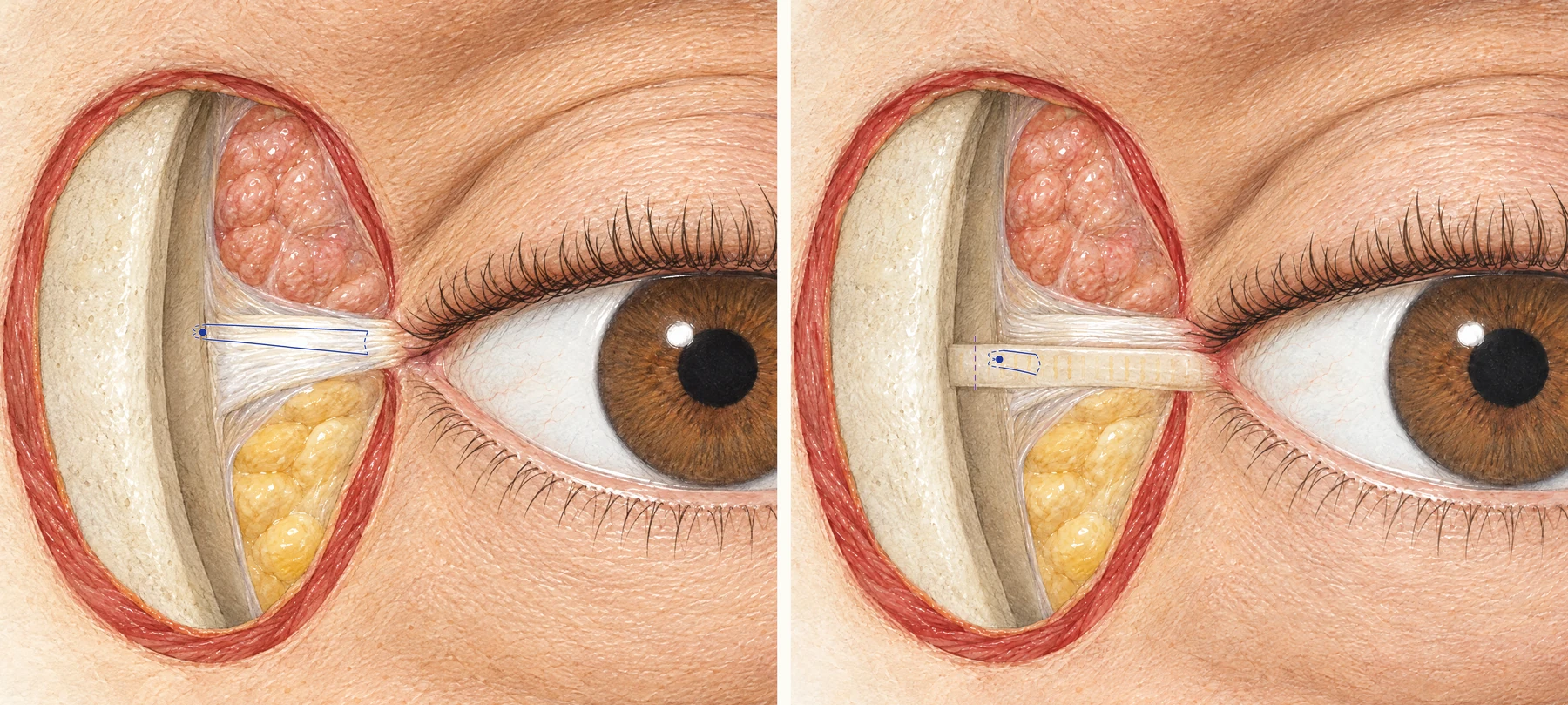
Canthopexy and canthoplasty
Two ways to tighten the outer corner of the eye. Left, canthopexy: the lateral canthal tendon stays attached and is stitched to the periosteum on the inner side of the bony orbital rim. Right, canthoplasty with a lateral tarsal strip: the corner is divided, the outer end of the lower eyelid tarsus is cut into a strip, shortened and stitched to the same spot inside the rim.
- Lateral orbital rim
- Inner surface of the orbital rim
- Lateral canthal tendon
- Suture from the tendon to the periosteum inside the rim
- Lacrimal gland
- Lateral fat pad, lower eyelid
- Orbicularis oculi, cut edge
- Lateral canthus
- Tarsal strip from the lower eyelid
- Upper limb of the lateral canthal tendon
- Trim line on the excess strip
- Suture from the strip to the periosteum inside the rim
- Canthotomy
Anatomy checked against StatPearls. Blepharoplasty, Lower Lid, Canthal Support, Anderson RL, Gordy DD. The tarsal strip procedure. Arch Ophthalmol 1979, Muzaffar AR et al. Surgical anatomy of the ligamentous attachments of the lower lid and lateral canthus. Plast Reconstr Surg 2002, StatPearls. Anatomy, Head and Neck: Eyelid.
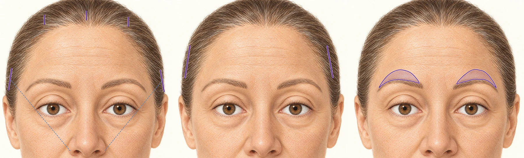
Brow lift incisions
Incision sites for three brow lifts. Left, endoscopic: three short cuts behind the frontal hairline and one behind each temporal hairline. Middle, temporal: one short cut in the hair of each temple. Right, direct: an ellipse of skin removed just above each eyebrow.
- Median incision
- Paramedian incision
- Frontal hairline
- Temporal incision
- Line from the nasal ala through the lateral canthus
- Temporal brow lift incision
- Temporal hairline
- Skin removed above the eyebrow
- Eyebrow
Anatomy checked against StatPearls. Endoscopic Brow Lift, Turin SY et al. Simplified Lateral Brow Lift under Local Anesthesia. Plast Reconstr Surg Glob Open 2019; Foustanos A. Lateral Brow Lift. Arch Plast Surg 2015, Perry JD, Hwang CJ. Direct Browlift. Clin Plast Surg 2022.
The operations
Each page covers what the operation treats, how it is done, the risks and the recovery, with the surgeons who show this work.